
Introduction
The original AREDS trial evaluated high-dose antioxidant vitamins consisting of vitamin C, vitamin E, and beta-carotene combined with high-dose zinc and copper.1 It demonstrated a 25% decrease in the five-year risk of progression to advanced AMD among high-risk patients.1 High risk was defined as intermediate AMD or noncentral GA in at least one eye, or advanced AMD in one eye and any stage in the fellow eye.1 This stage-based framework established oral supplementation as the first evidence-based intervention shown to modify the risk of progression to advanced AMD.1
AREDS2 refined the original formulation by replacing beta-carotene with lutein and zeaxanthin to improve safety, particularly for smokers due to the observed association between beta-carotene and lung cancer risk.2 Omega-3 fatty acids were also studied but demonstrated no added benefit and were not recommended for AMD risk reduction.2 The AREDS2 formula preserved efficacy while also improving safety.2 It remains a standard for risk reduction in appropriately selected patients with AMD.
It is also critical to clarify what AREDS2 does not do. Neither AREDS nor AREDS2 were designed to treat established GA or evaluate GA lesion growth as a primary outcome.1,2 AREDS2 Report 16 confirmed that supplementation does not slow GA enlargement once atrophy occurs, regardless of the AREDS2 treatment assignment.3 Assignment groups included the original AREDS formulation with the removal of beta carotene, a decreased zinc dosage or the addition of lutein/zeaxanthin, or omega-3 fatty acids.3 This has logically led some clinicians to question whether supplementation should be considered once GA is present.
POINT
Erika Anderson, OD, FAAO
AREDS2 CONTINUES TO HAVE A ROLE IN THE MANAGEMENT OF GEOGRAPHIC ATROPHY IN MANY PATIENTS
Building on the original Age-Related Eye Disease Study (AREDS), the Age-Related Eye Disease Study 2 (AREDS2) remains relevant for many patients with geographic atrophy (GA).1,2 This is not because it stops atrophy enlargement, but because it reduces the risk of progression from intermediate age-related macular degeneration (AMD), the Beckman clinical stage that most closely aligns with AREDS-defined treatment-eligible disease, to the advanced AMD, which includes the development of retinal neovascularization or center-involved GA.2 In clinical practice, GA rarely occurs in isolation. Many patients have asymmetric disease, where one eye has center-involved GA while the fellow eye remains at an AREDS2-eligible stage. Discontinuing supplementation simply because atrophy has occurred removes one of the few interventions supported by randomized trial data.1,2
AREDS2 should not be framed as a therapy to slow atrophy growth. Patients and clinicians are primarily focused on preserving function by delaying foveal involvement of GA, as well as reducing risk of conversion to neovascular disease. Enlargement is mainly driven by lesion characteristics such as baseline size, location, multifocal presentation, and bilaterality rather than supplement use.3 However, the risk of progression to advanced AMD remains clinically meaningful. In AREDS2, eyes that developed GA had approximately a 29% four-year risk of developing neovascular AMD.2,3 For many patients, the more immediate threat to vision is this conversion rather than atrophy expansion alone.3 Continuing AREDS2 is a practical way to manage this ongoing risk.
PREDICTING GA PROGRESSION: UNDERSTANDING DISEASE PROGRESSION AND THE USE OF OCT, FAF, AND EMERGING AI PREDICTION MODELS
Determining who may benefit from AREDS2 requires an individualized risk assessment. This involves establishing who is an appropriate candidate for therapy. Patients frequently ask, “When will I go blind?” Answering this question requires understanding predictors of progression and using newer tools to estimate prognosis and guide shared decision-making.
GA trials often use anatomic endpoints such as total lesion area and rate of enlargement because they allow for reproducible measurements and are sensitive to estimating treatment effects.4,5 Unfortunately, structural enlargement of GA does not completely correlate with real-world visual function.6,7 Vision is driven more by foveal involvement of GA and surrounding tissue integrity than total atrophic area.5,6 This structure vs function dissociation is central to the management of these patients. Enlargement is expected, but what matters most to patients is when the atrophy will reach the foveal center. Once foveal involvement occurs, eccentric fixation often develops. This impairs reading, driving, and other activities of daily living even when Snellen acuity appears preserved.5,6
Earlier prediction models relied on demographic, environmental, and genetic variables to estimate advanced AMD risk.8 Seddon et al showed that baseline AMD stage is the strongest predictor of advanced disease and developed a multivariable model that integrated smoking history, body mass index, and key genetic variants (CFH and ARMS2/HTRA1) into assessing risk.8 Systemic vascular health and medication exposures may also influence GA enlargement rates, suggesting progression is not driven solely by retinal features.9 These models helped at a population level but were less useful in predicting outcomes for an individual patient.
Modern GA care allows a more personalized approach using imaging available in routine practice. Optical coherence tomography (OCT) and fundus autofluorescence (FAF) biomarkers help estimate growth rate and foveal risk. Artificial Intelligence (AI) models, such as deep learning systems that predict progression from OCT and FAF imaging, are emerging as an additional tool in patient care.
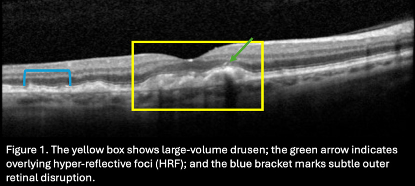
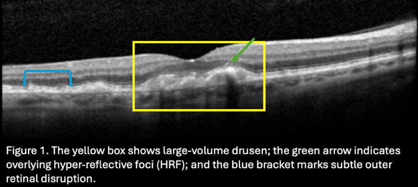
OCT can detect early structural changes that may proceed clinically apparent atrophy.10,11 Sleiman et al identified OCT features that were found to have a higher risk of progression to atrophic AMD, including hyperreflective foci, large or tall drusen, subretinal drusen deposits (reticular pseudodrusen), and disruption of the outer retinal layers including ellipsoid zone and photoreceptor changes.10 Several of these features are demonstrated in Figure 1.
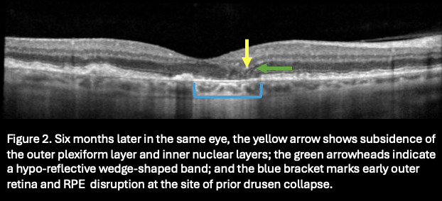
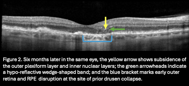
Nascent GA represents a preatrophy stage of AMD that can be identified on OCT before frank atrophy is visible on fundus examination.11 Key features include subsidence of the outer plexiform and inner nuclear layers, a hyporeflective wedge-shaped band, and early outer retinal and retinal pigment epithelium (RPE) disruption (Figure 2).11 These findings predict subsequent GA development.10 OCT biomarkers help distinguish eyes likely to remain stable from those at higher risk and can guide follow-up intervals and counseling.10,11
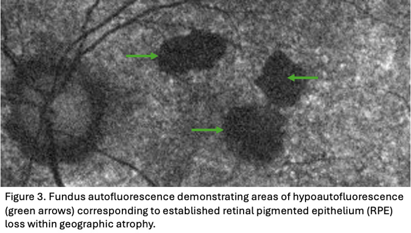
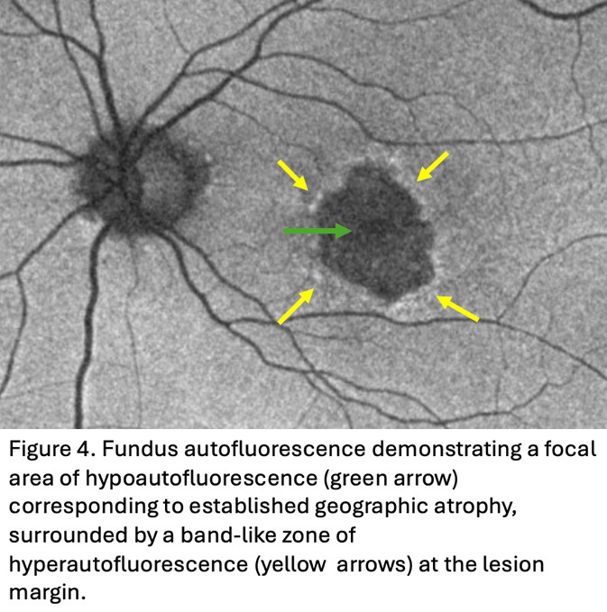
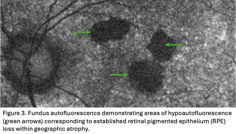
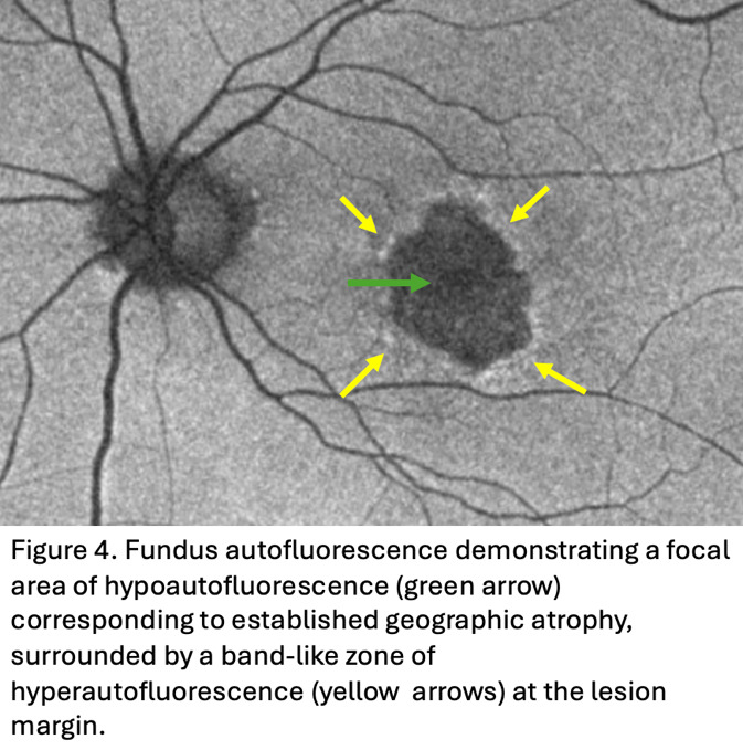
FAF complements OCT findings by highlighting established atrophy and adjacent RPE stress.12 Hypoautofluorescence corresponds to RPE loss, while hyperautofluorescence reflects metabolically stressed or “sick” RPE with lipofuscin accumulation (Figure 3).12 FAF abnormalities may precede clinically viable GA and help predict where atrophy will develop later.13 Junctional-zone patterns, particularly diffuse and banded patterns of increased autofluorescence, are associated with faster enlargement and greater foveal involvement risk (Figure 4).14 When FAF shows fovea-involving GA, AREDS2 is unlikely to benefit that eye, although the fellow eye may still qualify for supplementation consideration.3
Deep learning based AI models are increasingly being studied to predict GA onset and growth.15–17 These models analyze complex OCT and/or FAF patterns to generate patient-specific risk estimates.16,17 Some models can predict near term GA development from a single OCT scan, while multimodal systems combine OCT and FAF to forecast future lesion area and growth rate.15–17 These tools improve structural prediction, though most remain focused on anatomic rather than functional outcomes. Future integration of imaging with functional measures such as contrast sensitivity and reading performance may better estimate when structural progression converts to visual disability.15–17
GENETIC AND INDIVIDUALIZED DECISION-MAKING
Genetic variation in the complement pathway and the ARMS2/HTRA1 locus is strongly associated with AMD susceptibility.18,19 This has generated interest in whether having genotype information may help with risk stratification and aid in predicting progression to advanced AMD. This information can be beneficial to patient counseling and monitoring over the course of the disease. In the AREDS cohorts, risk variants in CFH and ARMS2/HTRA1 loci were also associated with a greater likelihood of progression to advanced AMD, supporting genotype as a contributor to baseline risk and the long-term disease course.18 Genotypes help predict who is more likely to develop advanced AMD but does not reliably predict how fast GA enlarges once atrophy is present.18,19 Near-term GA prognosis is better predicted by clinical findings (lesion size and foveal involvement) and imaging biomarkers (OCT and FAF lesion features) than by genotype alone.19
Awh et al raised interest in whether genotype influences response to zinc containing AREDS formulations.20 Released in 2015, the article proposed that supplementation efficacy may vary across CFH and ARMS2/HTRA1 risk profiles.20 This raised understandable questions about recommending AREDS2 supplementation without a patient’s genotype information. Subsequent studies and independent re-analyses have not confirmed clinically meaningful genotype treatment interactions.21,22 No prospective trials have demonstrated genotype specific harm from AREDS2.23 Therefore, genetic testing should not be used to withhold supplementation for patients in AMD stages where its benefit has been demonstrated.21–24
FUNCTIONAL OUTCOMES, PATIENT-CENTERED CARE, TREATMENT BURDEN, AND RISK TRADE-OFFS
Natural history studies have consistently shown that GA leads to progressive and irreversible functional vision loss.5,23,25,26 Patients with parafoveal disease often report trouble with reading, difficulty driving, and needing more light to read prior to a marked decrease in Snellen acuity.6,23 Standard visual acuity testing may underestimate a patients perceived visual disability.6 Functional measures such as reading speed and contrast sensitivity may be a better reflection of the patient’s visual limitations.6,23As lesions approach the fovea, acuity loss becomes more pronounced.6,25
Counseling in these patients remains essential. Smoking cessation is the most important modifiable risk factor for AMD progression and should be emphasized at every visit.22 Diet changes such as Mediterranean-style or plant-forward diets rich in leafy greens, legumes, whole grains, and sources of unsaturated fats are associated with a reduced risk of advanced AMD.22,27,28 Nutrient dense plant-based diets that include key micronutrients such as vitamin B12, omega-3 fatty acids, lutein, and zeaxanthin remain another reasonable dietary option.28 Photobiomodulation has also been explored, although its role in clinical care remains uncertain.
Intravitreal injections that inhibit the complement pathway represent an important advance in the care of patients with GA. Pegcetacoplan and avacincaptad pegol have been found to slow GA lesion enlargement when compared with sham with reductions in lesion growth rate of approximately 15-22% during their respective trials.29,30 The GALE extension study of OAKS and DERBY demonstrated increased treatment effect over time, with pegcetacoplan reducing GA growth up to 39% in monthly dosing and 32% with every-other-month dosing at 36 months, and 45% in nonsubfoveal GA.31 These therapies have not demonstrated meaningful improvements in visual acuity and require frequent ongoing intravitreal injections.29,30 Some risks associated with the treatments include conversion to neovascular AMD, intraocular inflammation or vasculitis, and injection related risk of endophthalmitis.29,30 For some patients, these trade-offs are acceptable while for others, the treatment burden and risks outweigh the potential benefit. This may be true in those with slower disease progression. In the future, investigational gene therapy for AMD may offer sustained intraocular delivery and reduce the need for frequent treatment.32 This could reduce treatment demands and expand available options to patients unwilling to commit to ongoing monthly injections.32
By comparison, AREDS2 supplements have a long-established safety profile and are generally well tolerated with gastrointestinal intolerance being the most common adverse effect.2 Although it is safe for most patients, clinicians should review a patient’s medical history, particularly the use of anticoagulant therapy, because the high-dosage of vitamin E may increase bleeding risk.22 Clinicians should avoid implying that AREDS2 slows GA lesion enlargement. Instead, they should frame it as a stage-appropriate strategy to reduce the risk of progression to advanced disease.2,3,22 In patients with asymmetric or nonfoveal involvement, or those hesitant to pursue invasive injection therapy, supplementation combined with lifestyle modification offers a practical, low-risk way to remain proactive while also keeping expectations aligned with what the current evidence can deliver.
COUNTERPOINT
Kensington Hatcher, OD, FAAO, FORS
ROUTINE AREDS2 SUPPLEMENTATION IS NOT NECESSARY IN PATIENTS WITH ESTABLISHED GA
For many clinicians, recommending AREDS or AREDS2 vitamins to patients with AMD has become almost reflexive. The formulations have been part of AMD management for more than 20 years, and for good reason: they reduce the risk of progression from intermediate AMD to advanced disease, particularly neovascular AMD.1 What is far less clear—and much less often questioned—is whether that same recommendation makes sense once a patient has already developed GA.
As new therapies for GA become available, and the way we talk to patients about this disease evolves, it is worth revisiting whether routine AREDS supplementation in this population is truly evidence-based or simply habitual.
AREDS WAS DESIGNED FOR PREVENTION, NOT TREATMENT OF ESTABLISHED GA
The most important point is also the simplest: AREDS was not designed to treat GA. Neither the original AREDS trial nor AREDS2 evaluated AREDS vitamins as a treatment for established GA.1,2 Both studies focused on whether supplementation could reduce progression from earlier stages of AMD to advanced disease, defined as either neovascular AMD or central GA. While the results supported use in intermediate AMD, they did not show that AREDS slowed GA enlargement, preserved vision once GA was present, or altered the natural course of the disease.1,2
Despite this, AREDS is still commonly recommended to patients who already have GA, often without a clear explanation of what benefit (if any) is expected. This blurring of prevention and treatment is understandable, but it matters. Preventing disease is not the same as treating established pathology, and the evidence supporting one does not automatically justify the other.
FUNCTIONAL OUTCOMES DO NOT IMPROVE WITH AREDS IN GA PATIENTS
From the patient’s perspective, GA is primarily a functional disease. Patients care about reading, driving, recognizing faces, and maintaining independence. Unfortunately, AREDS supplementation has not been shown to improve any of these outcomes in patients with GA.1,2 Natural history studies have consistently demonstrated that GA lesions enlarge at a relatively steady rate over time and that rate appears largely unaffected by antioxidant supplementation.4,5 Visual acuity and functional vision decline as atrophy progresses, regardless of whether a patient is taking AREDS.
When clinicians tell GA patients that AREDS may “slow progression,” it is worth asking what that statement is based on. If lesion growth, visual function, and patient experience remain unchanged, the clinical relevance of continued supplementation becomes difficult to justify.
DISEASE PROGRESSION CONTINUES DESPITE SUPPLEMENTATION
GA is characterized by a gradual and largely predictable enlargement of atrophic lesions over time, with associated decline in visual function as central involvement progresses. Multiple longitudinal studies have demonstrated relatively consistent rates of GA enlargement, with average lesion growth reported at approximately 1.5 to 2.1 mm2 per year, although variability does exist between individuals and lesion patterns.3,4 Importantly, AREDS and AREDS2 were not designed to modify the course of established atrophic disease and several observational studies have also shown that GA progresses predictably whether or not patients are taking AREDS.33
Some clinicians point to a possible reduction in neovascular conversion, particularly in the fellow eye, as a reason to continue supplementation. While that argument may apply in select cases, it does not support a blanket recommendation for all GA patients. Many individuals already have bilateral advanced disease, leaving little theoretical benefit to be gained from ongoing supplementation.
TREATMENT BURDEN IS OFTEN OVERLOOKED
AREDS vitamins are often described as low risk, but “low risk” is not the same as no burden. Patients are typically advised to take these supplements indefinitely, resulting in ongoing costs that can be meaningful for older adults on fixed incomes. Pill burden also matters, particularly in patients already managing multiple systemic medications. In a busy clinic, time spent reinforcing a supplement with limited benefit may come at the expense of discussions that are far more impactful such as low-vision referral, home safety, assistive technology, or long-term planning.
Adverse effects are uncommon, but they are not nonexistent. High-dose zinc has been associated with increased genitourinary hospitalizations, and some patients experience gastrointestinal side effects or intolerance.1 When the expected benefit is minimal, even small risks take on greater importance in the overall risk–benefit calculation.
THE MANAGMENT PARADIGM FOR GA IS CHANGING
The treatment landscape for GA is also changing. Until recently, management largely consisted of monitoring progression and helping patients adapt to vision loss. With the introduction of FDA-approved complement inhibitors such as pegcetacoplan and avacincaptad pegol, GA is no longer a condition managed solely with observation and reassurance.29,30 These therapies are not without risk and require careful patient counseling, but they represent true attempts to modify disease progression—something nutritional supplementation has not been shown to do. Continuing to emphasize AREDS as a default recommendation risks anchoring both clinicians and patients to an outdated management paradigm.
INDIVIDUALIZED COUNSELING IS PREFERABLE TO ROUTINE RECOMMENDATION
None of this is meant to suggest that AREDS should never be discussed with patients who have GA. There are reasonable scenarios where supplementation may still be considered, such as patients with GA in one eye and intermediate AMD in the fellow eye, or patients who prefer to continue vitamins after a clear discussion of the limitations. What is difficult to justify, however, is the routine, unqualified recommendation of AREDS for every patient with GA.
There is also an ethical component to consider. When clinicians recommend a therapy, patients assume it is expected to help. If a patient with GA is told to take AREDS and then experiences the expected progression of disease, disappointment and confusion are understandable. Clear communication about what AREDS can and cannot do is essential so patients can make informed choices.
SUMMARY
AREDS2 supplementation has an established role in the management of selected patients with AMD, particularly those with intermediate disease. However, its role in patients with established GA is less clear. In individuals with advanced bilateral or foveal-involving GA, the available evidence suggests minimal impact on disease progression or visual outcomes, making routine supplementation difficult to justify.
The AREDS2 formulation was developed to target oxidative stress, a central pathway in AMD pathophysiology. However, growing evidence suggests oxidative injury alone does not fully explain disease advancement.34 A recent review in Ophthalmology and Therapy summarized observational findings from the AREDS and AREDS2 cohorts showing that higher dietary intake of vitamins B6, B9, and B12 were associated with lower rates of progression to advanced AMD.34 This may be related to homocysteine-mediated vascular and inflammatory effects within the retina, processes influenced by B-vitamin metabolism.34 Because these pathways are not directly addressed by the current AREDS2 formulation, there is a growing interest into whether targeted B-vitamin supplementation could complement existing antioxidant strategies by addressing additional mechanisms involved in disease progression.34 This is currently being marketed as AREDS3, although no large randomized study has been completed.
In the end, supplementation decisions should reflect disease stage, fellow-eye status, and patient priorities, with additional considerations such as cost and pill burden. Therapeutic options for GA remain limited and no intervention restores vision that is already lost. Although the treatment landscape is evolving, GA remains a chronic, relentless, and degenerative condition, and no therapy can fully stop it. Until treatments can preserve and restore function, thoughtful counseling and shared decision-making remain central to patient care.
Conflicts of Interest
KH: Employee of Neurotech
Funding Sources
None