
INTRODUCTION
When 2 dissimilar visual stimuli are presented to each eye, the visual system may adapt by suppressing input from one eye to avoid perceptual conflict. In children with poorly controlled intermittent exotropia, this sensory adaptation often occurs, preventing diplopia.1,2 Current nonsurgical therapies, including patching3 and binocular vision training,4,5 have demonstrated success in improving control of exodeviation. One of the therapeutic goals in these approaches is to remediate suppression, as persistent suppression may hinder the recovery of fusion and binocular stability.
Despite the importance of managing suppression in exotropia patients, there remains a lack of simple clinical tools that allow clinicians to modulate suppression in a graded manner during office-based therapy. Standard tests, such as the Worth 4-dot test, red lens test, and Bagolini test, are well-established clinically for detecting suppression, yet they offer limited insight into its depth. When encountering deep suppression, clinicians often toggle room illumination to break suppression. However, these methods only yield a binary suppression status (present/absent) under either bright or dim lighting conditions.6 Moreover, the abrupt luminance shift may unintentionally reduce latency and lead to an inaccurate overestimation of binocular stability.7 To better estimate the suppression severity, specialized filters, such as 10-step neutral-density filters8 and Sbisa bars,9 are reportedly utilized in discrete steps rather than along a continuous scale.
When managing patients with manifest exotropia, pathological diplopia awareness training is important. In practice, however, this often requires simultaneous adjustments to several parameters, including room illumination, the use of filters, and changes in target size or contrast. A simple clinical tool that could both alter interocular luminance in a wide continuous range and directly regulate its level during therapy would be very desirable. Such an approach could streamline therapeutic management and potentially make vision training more efficient for patients with deep suppression.
This case report describes the novel use of a variable neutral-density filter as both a diagnostic and therapeutic tool for vision training in a child with intermittent exotropia and deep suppression. By progressively reducing the luminance reaching the dominant eye, the filter can lessen interocular imbalance and enhance binocular participation during both assessment and training. This approach facilitated the vision training process, with clinically and subjectively satisfactory ocular alignment for both the patient and the parents, achieved after 12 sessions—fewer than the more typical 20–25 sessions reported in office-based vision therapy.10
CASE REPORT
Initial Visit
An 8-year-old girl presented to the binocular vision clinic with a history of worsening left eye exotropia over the past 6 months. According to her parents, they had first noticed an outward deviation of the left eye at approximately 3–4 years of age, during which time the eye was reportedly deviated for 70%–80% of waking hours. She had previously been evaluated by both ophthalmologists and optometrists; however, no treatment was initiated because they reported that amblyopia was not present.
Her parents reported that she was born full-term via cesarean delivery, with unremarkable systemic and developmental histories. There was no history of long-term medication use or prior ocular surgery. However, regarding the recent increase in the magnitude of the exodeviation, particularly over the preceding months, her parents were concerned about cosmetic appearance and sought consultation regarding the potential role of vision therapy.
The unaided entrance visual acuity was 20/20 in each eye. Worth 4-dot testing demonstrated suppression in the left eye across all distances and lighting conditions. Pupils were equal, round, reactive, and had no relative afferent pupillary defect. Extraocular motilities and confrontation fields were full in both eyes and no A/V pattern was found. Retinoscopy and subjective refraction revealed right eye +0.25 and left eye plano. The binocular vision evaluation revealed constant left exotropia at both distance and near, graded by the Intermittent Exotropia Control Score (level 5).11 An alternating cover test, neutralized by the Fresnel prism over the subjective refraction, showed 50∆ exotropia at distance and 55∆ exotropia at near. The synoptophore (first-degree targets Ex and square were used) confirmed equality of the subjective and objective angles at 50∆ Base-in, indicating normal retinal correspondence.
The monocular accommodation amplitudes of the right and left eyes were 20 D. Accommodation facility with ±2.00 D was 18 cycles per minute in the right eye and 16 cycles per minute in the left eye. No binocular accommodative facility was attempted due to manifest exotropia. The calculated accommodation-convergence/accommodation ratio was 3.75/1.
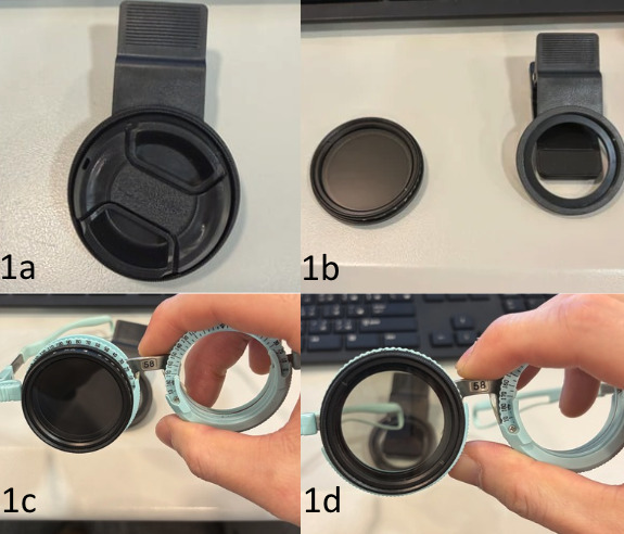
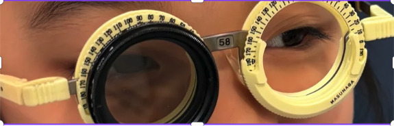
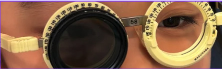
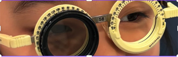
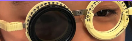
Even with an additional −3.00 D lens at near, she was unable to achieve gross convergence at near. Stereopsis was further evaluated using the Random Dot 2-S Stereo Acuity Test (Vision Assessment Corporation, United States) in conjunction with the −3.00 D lens. However, no local and global stereopsis could be elicited. In addition to conventional binocular vision testing, a variable neutral-density filter (Shenzhen Shixin Trading Company, China) was used to assess the depth of suppression (Figure 1).
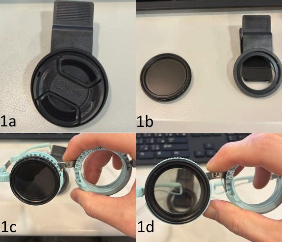
Variable neutral-density filters used by professional photographers consist of 2 stacked linear polarizers. The reduction in luminance when one polarizer rotates relative to the other is governed by Malus’ Law:
I=I0cos2(θ)
Where:
-
= The resultant intensity of light passing through the polarizers.
-
= The initial intensity of light passing through the first polarizer.
-
θ = The angle between the transmission axes of the 2 polarizers.
In optometry, filter strength is often expressed as optical density, the negative logarithm of transmittance. Transmittance is defined as
By substituting Malus’ Law into the density formula, the following table provides a conversion from the angle of rotation in the trial frame to the equivalent optical density used in a clinical setting (Table 1). The values reported in Table 1 are at discrete 10-degree increments to demonstrate the progression of transmittance or optical density. However, the filter can be adjusted to present any degree between 0 and 90.
Given that the patient exhibited constant suppression on previous Worth 4-dot testing, a variable neutral-density filter was placed in front of the fixating right eye, and the test was re-performed. In the presence of manifest exotropia, she was asked to report the number of dots perceived at 40 cm with the light off. The optical density of the filter was then gradually increased. At 60° of rotation (approximately 0.6 log unit of attenuation), she reported pathological diplopia (perception of crossed 5 dots).
To further evaluate if stereopsis could be elicited under altered interocular luminance conditions, a −3.00 D lens was placed in front of both eyes, and the filter was rotated to 60° in front of the right eye. Under these conditions, the near point of convergence improved to 20 cm. The patient was able to perceive the random-dot stereoscopic target, with a base-out fusional reserve of 8∆ and a base-in reserve of 1.5∆ in the computerized dichoptic training program at 40 cm, indicating a favorable prognosis for vision therapy.
The incorporation of the variable filter demonstrated that the suppression in this case was not absolute. Under specifically modified visual conditions, the deviation exhibited intermittent features, and the patient was able to re-establish fusion. After discussion, she was diagnosed to have basic type, large, constant left exotropia (Intermittent Exotropia Control Score: Level 5 at both distance and near). In view of the substantial magnitude and deep suppression, both strabismic surgery and vision training were discussed. However, parents preferred a nonsurgical approach. Vision therapy was then discussed, and a 3-month training program was recommended to evaluate whether training alone could produce meaningful improvements in ocular alignment and cosmetic appearance. The patient was asked to schedule sessions every 1–2 weeks. Daily 15–30 minutes of home vision therapy was recommended. Parents and the patient were informed that approximately 20–25 lessons might be required.
VISION THERAPY SESSIONS
A total of 12 vision therapy sessions were completed, which was significantly fewer than planned. The 12-session vision therapy protocol (Table 2) targeted monocular skills (accommodation, pursuits, and saccades), anti-suppression, and vergence. Treatment began by normalizing accommodation facility, awareness, and saccadic eye movement, progressing from gross to fine saccades. To eliminate deep suppression, the variable neutral-density filter was first placed over the dominant eye during binocular training, including the Brock String, cheiroscope, aperture rule, anti-suppression Hart Chart reading, eccentric circles, and free-space fusion with random-dot stereograms. The density of the neutral-density filter was gradually increased until suppression was eliminated. As the interocular sensory balance improved throughout the training, the filter was subsequently removed and then applied to the nondominant eye. This intentional sensory stress introduced an additional binocular challenge in maintaining alignment. Training advanced from near to distance, from smooth to step vergence. Ultimately, all visual skills were integrated. Both the patient and parents reported excellent adherence to the home reinforcement regimen. The patient was motivated by her own observation of improved ocular alignment, which encouraged consistent engagement in the prescribed 30-minute daily training sessions throughout the treatment course.
Outcome
After 12 vision therapy sessions, the patient was exophoric. Both the parents and the patient expressed satisfaction with the cosmetic outcome, noting that her eyes appeared straight most of the time, with manifest exotropia occurring about once per week. On occasions when exotropia decompensated during fatigue, the patient reported that she could voluntarily regain fusion with minimal effort. Her entering distance and near visual acuities were 20/20 in each eye, and subjective refraction was unchanged. Clinical evaluation findings from the initial visit, discharge, and the 4-month follow-up are compared in Table 3. Although residual intermittent exotropia persisted, the patient’s office control score improved markedly from level 5 to level 1, a measure recognized in previous literature as a primary indicator of clinical success.4,5 A reduction in the exodeviation magnitude was also noted (from 50∆ to 30∆ at distance and from 55∆ to 30-35∆ at near), as shown in previous literature.12 This ≥10∆ change can be regarded as a significant change rather than a measurement error,12 and may be attributed to the improved tenacious proximal fusion13 or slow fusional vergence.14,15 When integrated with objective improvements in near point of convergence, accommodation facility, sensory-motor fusion, alongside positive subjective reports of ocular alignment from both the patient and parents, this case was considered clinically cured.
At discharge, the patient was prescribed home maintenance therapy, including eccentric circles at far and near, together with lens rock with flippers. She was instructed to perform home therapy twice weekly and to return in 3 months for reassessment. At the 4-month progress evaluation, the patient and parents reported good control of eye alignment. No suppression was found in the Worth 4-dot testing at all distances under standard room illumination. Other clinical findings remained unchanged. After discussion, the patient chose to continue home-based exercises 1–2 times per week, reporting that they were easy to perform and were not burdensome.
DISCUSSION
This case highlights a novel clinical application of a commercially available variable neutral-density filter in the management of poorly controlled exotropia (control score of 5) with deep suppression. Although discrete 10-step neutral-density filters8 or Sbisa bars9 can be utilized to attenuate interocular luminance and quantify suppression, their fixed-step nature can limit precise measurement. In contrast, the variable neutral-density filter used in this case report provides a continuous, gradual assessment of suppression depth. By combining the variable filter with the Worth 4-dot test, the patient’s scotoma depth can be evaluated, and the precise luminance transmittance required to eliminate suppression can be recorded. This offers a more accurate and objective measure of sensory improvement across visits than conventional suppression testing.
Another notable advantage is that a gradual reduction of luminance to the dominant eye may allow convergence training to begin earlier than would be possible using standard methods alone. In patients with significant exodeviation and suppression, direct binocular training is often delayed because the patient cannot appreciate simultaneous perception or fusion under standard viewing conditions. Although over-minus power or base-in prism may be used to reduce the angle of deviation for training, these methods may not be useful in patients with exodeviation of 40∆ or more.6 As a result, therapy may need to begin with prolonged monocular or synoptophore activities before meaningful convergence tasks can be introduced. In the present case, reducing the luminance of the dominant eye enabled testing of gross convergence and training at an earlier stage. Earlier access to binocular tasks may, in turn, shorten the overall duration of therapy by accelerating the transition from sensory preparation to active vergence rehabilitation. Furthermore, the variable filter transformed into an active sensory challenge during the final stages of training. By placing the filter over the originally deviated eye rather than the dominant eye (Table 2), this added additional sensory loading and challenged the stability of the patient’s binocular system. This dual capability highlights the variable filter’s rehabilitative utility in this case.
The gradual luminance modulating feature also has important implications for task simplification in vision therapy. Conventionally, trainings for intermittent exotropia and associated suppression often require clinicians to modify target characteristics—such as size, brightness, contrast, detail, color, or contour similarity—to make binocular integration more achievable. Although this approach is well-established, not all target parameters can be easily manipulated using available office- or home-based therapy tools. In addition, assembling multiple tools with a wide range of visual characteristics may impose a considerable financial burden on both practitioners and patients. This is particularly relevant in settings where access to specialized computerized systems or an extensive range of vision therapy materials is limited. The variable neutral-density filter offers a comparatively low-cost alternative (approximately $5 USD). Instead of manipulating multiple instruments to alter the target content, the therapists can readily modify the luminance reaching the dominant eye while keeping the same training target and task structure. This simplifies the rehabilitation process and allows suppression to be challenged in a graded, reproducible fashion. The level of suppression can be adjusted and recorded systematically, making it possible to monitor sensory improvement objectively across visits.
Despite these advantages, the use of a variable neutral-density filter should be interpreted as an adjunct rather than a replacement for established binocular vision assessment and therapy techniques. Further studies would be valuable to determine its reproducibility, its reliability compared to other established suppression grading methods, and its effectiveness across different types and severities of strabismus. In addition, its use should only be limited to in-office training, as the filter’s luminance reduction depends on the absolute luminance.16 Given that ambient luminance can vary substantially over the course of a day, in some circumstances by as much as 100000:1,16 the therapeutic effect may be less predictable unless lighting conditions are carefully controlled (for example, in a clinic cubicle). Nonetheless, this case report demonstrated the meaningful clinical utility of the filter. It offers a simple means of continuously grading suppression, reduces dependence on multiple specialized therapy tools, and facilitates the timely initiation of convergence training by progressively reducing luminance to the dominant eye. Together, these features support its potential role as both a diagnostic and therapeutic instrument in the management of exotropia with deep suppression.
CONCLUSION
This case report illustrated the usefulness of a variable neutral-density filter as an inexpensive adjunct in the management of exotropia by permitting continuous grading of suppression and accelerating sensory rebalancing during vision therapy, ultimately contributing to a shorter treatment course. Although further study is obligatory, this low-cost approach has practical diagnostic and therapeutic insights in selected cases of exotropia with significant suppression.
TAKE HOME POINTS
-
A variable neutral-density filter can provide a simple and continuous method for grading suppression.
-
The filter may function as a therapeutic tool by reducing luminance to the dominant eye, thereby rebalancing interocular sensory input and promoting binocular participation.
-
In patients with large-angle exotropia and deep suppression, gradual luminance attenuation may allow earlier initiation of convergence training, potentially shortening the overall duration of therapy.
Patient Consent
Consent was granted by the patient and parents for the publication of this case report and any accompanying clinical images.
Human Rights
Procedures were followed in accordance with the Helsinki Declaration. A case report of 3 or fewer patients does not require institutional review board approval.
Conflicts of Interest
The author declares no conflicts of interest.
Funding Sources
The author declares no funding sources.
Data Availability
Data sharing is not applicable to this manuscript.
Commercial Sponsorship
The author declares no commercial sponsorship received.
Artificial Intelligence (AI) use
AI was not used in the creation or submission of this manuscript.