
INTRODUCTION
Papilledema is defined as bilateral optic nerve head edema caused by increased intracranial pressure.1 This increase in intracranial pressure can be caused by space-occupying lesions, infections, or trauma, among other etiologies.1 Papilledema may also be induced from iatrogenic reasons, as some medications can increase intracranial pressure.2 More often than not, there is no known etiology of the patient’s increased intracranial pressure and subsequent optic nerve head edema, leading to the diagnosis of idiopathic intracranial hypertension, a diagnosis of exclusion.3 Patients with papilledema may present with symptoms of headaches, pulsatile tinnitus, nausea, double vision or vision changes, such as vision loss or transient visual obscurations, among other symptoms.1
Drug-induced papilledema is a known side effect of certain drug types or classes, the most common being tetracyclines or vitamin A derivatives.2 Leuprolide acetate (“Lupron”, AbbVie Endocrine Inc., North Chicago, IL) is a synthetic gonadotropin-releasing hormone agonist with various clinical indications, such as treating fibroids in endometriosis in women, prostate cancer in men, central precocious puberty in children, and in vitro fertilization to list a few.4 While papilledema has been reported with leuprolide acetate use, such published cases remain exceedingly rare (Table 1).5–9
This report highlights the presentation of leuprolide acetate induced papilledema in a young female patient soon after starting the medication to prepare for a hysterectomy. The pathophysiology, treatment, and management are reviewed.
CASE REPORT
A 35-year-old Black female presented to the eye clinic after being diagnosed with bilateral optic disc edema of unknown origin at an outside clinic. The patient was examined by the outside clinic 15 days earlier and had recommended the patient self-present to the emergency department, but the patient did not seek emergent care. The patient noted sinus pressure temporally and photophobia but denied blurry vision, pulsatile tinnitus, or diplopia. The patient had a notable medical history of uterine fibroids with a planned hysterectomy in one month. To prepare for the surgery, the patient was prescribed an unknown dosage of monthly leuprolide acetate injections, of which she was currently on the fifth month of her 6 total months of treatment. Two weeks prior to beginning leuprolide acetate injections, the patient received 2 iron infusions each one week apart to treat iron deficiency anemia. After beginning leuprolide acetate injections, the patient noted rapid weight gain of about 60 pounds resulting in a current body mass index (BMI) of 32.4 (Class I obesity).10 Since the patient began the medication, she noted systemic side effects of the drug including hot flashes, intermittent headaches, and intermenstrual bleeding. The patient had no other medications and no known medication allergies. Her ocular history included being diagnosed in 2016 with pseudo-papilledema of both optic nerves, for which she was previously followed at the clinic with optical coherence tomography imaging. Her left eye had myelination of the retinal nerve fiber layer superior to the optic nerve.
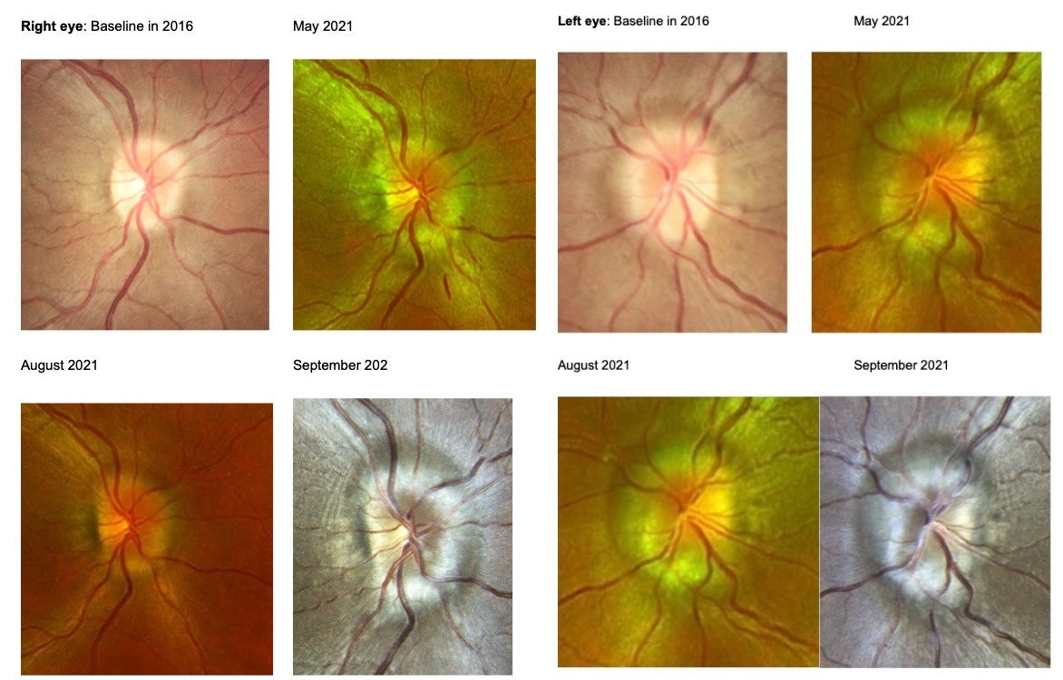
Best corrected visual acuity was 20/20 in the right eye and the left eye. Extraocular motilities and confrontation visual fields were full. Pupils were equal, round, and reactive to light with no afferent pupillary defect of either eye. Slit lamp evaluation was remarkable for trace papillae of the conjunctiva and capped meibomian glands in both eyes, suggestive of allergic conjunctivitis and mild meibomian gland dysfunction, respectively. Intraocular pressures were measured as 17 mmHg in the right eye and 15 mmHg in the left eye with Goldmann Applanation Tonometry. The patient’s blood pressure measured 142/85 mmHg. Dilated fundus examination revealed edematous and elevated optic nerve heads with indistinct margins and obscured blood vessels in both eyes (Figure 1). There was a notable inferior flame hemorrhage in the right eye and no visible hemorrhages in the left eye. The patient’s optic nerve head edema was consistent with grade III papilledema in both eyes on the Frisén scale. All other dilated exam findings were unremarkable.
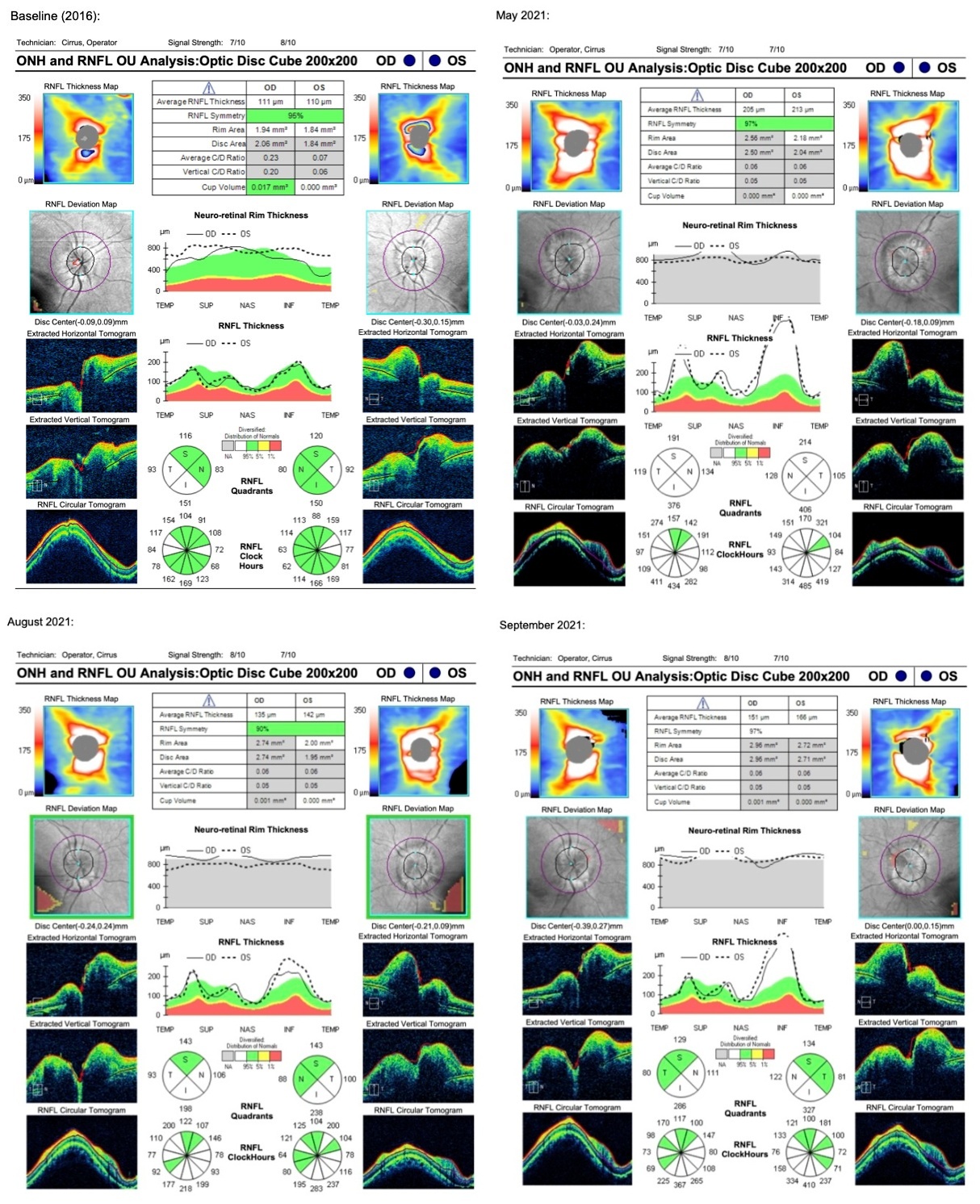
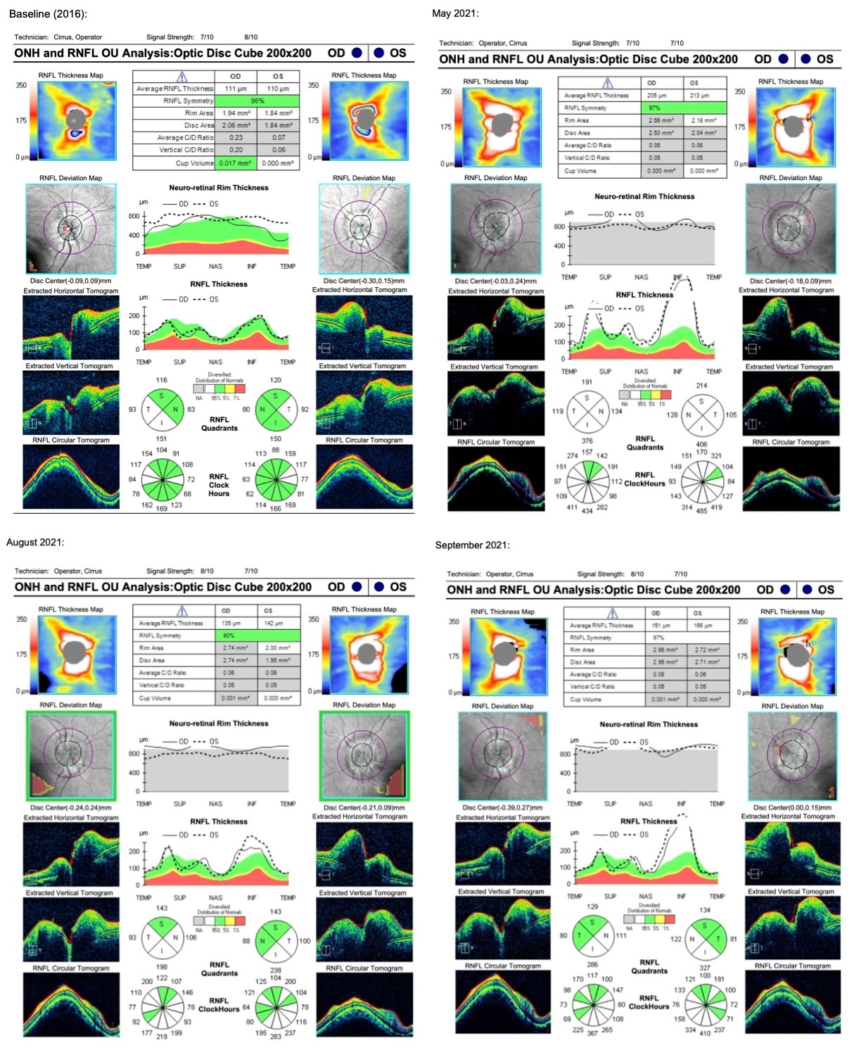
As the patient was previously monitored for pseudo-papilledema, a previous baseline optical coherence tomography 5 years prior showed an average retinal nerve fiber layer thickness of 111 microns and 110 microns in the right and left eye, respectively (Figure 2). Retinal nerve fiber layer optical coherence tomography was repeated at this visit showing an increase in nerve fiber thickness to 205 microns in the right eye (94 micron increase from baseline), and 213 microns in the left eye (103 micron increase from baseline). The concern for drug-induced papilledema was considered after reviewing the patient’s history, current medication, and examination findings. The patient was previously told by the outside provider to self-present to the emergency department and this was restressed at the exam, but the patient was averse to presenting to the emergency room. It was then recommended that the patient see her primary care provider urgently to undergo neuroimaging due to concern for possible papilledema. One week later, there was an attempt to contact the patient via telephone to ensure her follow-up with primary care, but the patient was unable to be reached.
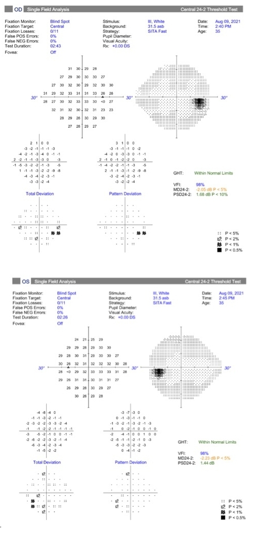
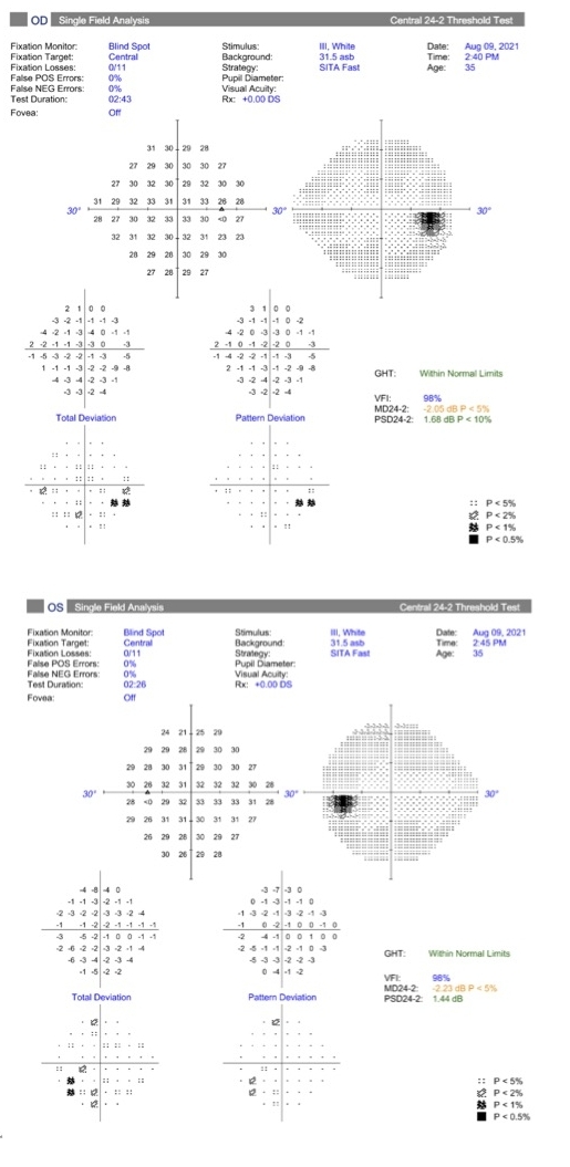
Three months later, the patient presented for a follow-up and reported she had undergone the scheduled hysterectomy without complication and had since discontinued leuprolide acetate injections following the sixth and final injection. The patient had not been prescribed any new medications and had not undergone the previously recommended neuroimaging. The patient’s calculated BMI was slightly lower than the initial visit, 30.5 (Class I obesity), and denied any current symptoms of headaches or pulsatile tinnitus following her hysterectomy 2 months prior.10 Visual acuity was stable at 20/20 in the right and left eye with stable afferent and efferent testing and slit lamp examination. The patient’s intraocular pressure was 18 mmHg in the right eye and 15 mmHg in the left eye. Upon dilation, the optic nerves remained mildly edematous but had improved from the previous visit, a grade II on the Frisen scale (Figure 1). Repeat optical coherence tomography imaging was performed, confirming a decrease in retinal nerve fiber layer thickness, with an average of 135 microns in the right eye (decrease of 70 microns) and 142 microns in the left eye (decrease of 71 microns) (Figure 2). A 24-2 Humprey Visual Field was performed at this visit which was relatively reliable and showed an essentially normal visual field of the right and left eye (Figure 3). The patient was strongly advised to undergo neuroimaging at this time.
_at.jpg)
Magnetic resonance imaging of brain and orbits was obtained along with magnetic resonance venography 2 weeks later. The magnetic resonance imaging revealed mild prominence of bilateral optic nerve sheaths and thickening at the bilateral optic nerve insertions, along with a partially empty sella. The magnetic resonance venography showed moderate to severe narrowing of junctions of the transverse and sigmoid sinuses. The imaging was suggestive of increased intracranial pressure leading to optic nerve head edema. It was recommended that the patient undergo a lumbar puncture, which she declined, as she preferred to lose weight prior to obtaining the lumbar puncture. After imaging was obtained, the patient was followed by neurology at an outside hospital.
The patient returned 6 weeks later for a comprehensive eye exam and denied any symptoms such as headaches or pulsatile tinnitus. The patient’s corrected entering visual acuity was measured at 20/20 in the right and left eye. The patient’s BMI at this visit was 31.52 (Class I obesity), slightly higher than her weight recorded 6 weeks prior.10 The patient’s dilated fundus exam revealed blurred optic nerve head margins with mild folds and vessel obscuration, a grade III on the Frisén scale (Figure 1). The optic nerve head optical coherence tomography was repeated, and the average retinal nerve fiber layer thickness was shown to have increased to 151 microns in the right eye (increase of 16 microns from previous visit) and 166 microns in the left eye (increase of 24 microns from previous visit) (Figure 2). Since the patient was also being closely followed by neurology, a follow-up was scheduled in 1 month to repeat the visual field; however, the patient was lost to follow-up.
DISCUSSION
The body’s natural gonadotropin-releasing hormone is produced in the hypothalamus and has different functions in male and female bodies.11 In those assigned a female sex at birth, gonadotropin-releasing hormone stimulates gonadotropin-releasing hormone receptors in the pituitary gland to make and release follicle-stimulating hormone and luteinizing hormone.11 The produced hormones ultimately affect sex steroid hormone production, such as estrogen and progesterone.11 Leuprolide acetate is a long-acting, synthetic gonadotropin-releasing hormone super agonist that is more potent than naturally occurring gonadotropin-releasing hormone.4,12,13 The half-life of endogenous gonadotropin-releasing hormone is 3 to 4 minutes, while the half-life of leuprolide acetate is about 3 hours.4 The drug mimics an excessive amount of gonadotropin-releasing hormone, initially spiking sex hormones.14 However, consistent overstimulation of the receptors causes a downregulation of follicle-stimulating hormone and luteinizing hormone with time.14 Ultimately, this causes a decrease in sex steroid hormone production, the therapeutic outcome for which leuprolide acetate is typically prescribed, which is completely reversible when discontinuing the medication.4
Leuprolide acetate may be prescribed as a monthly injection for those with endometriosis or as hormonal regulation treatment for uterine fibroids.4 Other indications for leuprolide acetate use are prostate cancer, central precocious puberty, breast cancer, and in vitro fertilization.4,14 Fibroids, also known as myomas, are benign tumors within the uterus and occur in about one fourth of women over the age of 35.4,15 Since these tumors directly respond to sex steroid hormones, the down regulation of hormones such as estradiol and progesterone with leuprolide acetate usage cause these tumors to shrink.4 Leuprolide acetate is a temporary treatment for fibroid reduction and may be prescribed in addition to iron to achieve the desired result of shrinking fibroids and reduce bleeding during a surgical hysterectomy, as seen in the patient’s case.15,16
Gonadotropin-releasing hormone receptors are found on membranes of pituitary gonadotrophs but are also found widespread throughout the body, such as on the liver, heart, and skeletal muscles.4,17 Since receptors for gonadotropin-releasing hormone are found in many areas of the body and change hormone levels in patients, many systemic side effects have been reported with the drug’s usage.4 Overall, leuprolide acetate is known to be a safe drug, with side effects being variable based on dosage, and mirror menopausal-like symptoms in female patients, such as night sweats, hot flashes, reduced libido, weight changes, changes in menses, and bone density loss.4 While our patient was using leuprolide acetate, her systemic side effects aligned with those in the literature including hot flashes, intermittent headaches, and intermenstrual bleeding. Leuprolide acetate usage has also been linked to hypertension and thrombosis in adults.18,19 The most common ocular side effects of leuprolide acetate include blurred vision, both transient after injection and long-term, eye pain, and eyelid edema.20 Rare ocular side effects include retinal hemorrhage or vein occlusion.19 Research in teleost fish revealed certain retinal cell types present in the retina are affected by gonadotropin-releasing hormone.21 The 2 areas of the retina affected were the inner nuclear layer and ganglion cell layer, which may provide insight as to how retinal side effects can occur with these medications.21
Another rare ocular side effect of leuprolide acetate usage is papilledema.5 Papilledema is defined as bilateral optic nerve head edema due to an increase of intracranial pressure.1,22 Patients may have an increase of intracranial pressure without papilledema, but by definition, papilledema is due to an increase of intracranial pressure.1 A patient is diagnosed with idiopathic intracranial hypertension, also known as pseudotumor cerebri, if there is optic nerve head edema and an increase in intracranial pressure without an identifiable etiology.1,23 This common diagnosis of papilledema is most frequently encountered in female patients who are obese, of child-bearing age, and under 45 years old but may be diagnosed in any age, or sex assigned at birth.1,23,24 Other causes of papilledema include but are not limited to space-occupying lesions, infections, or trauma.1 Iron deficiency anemia has also been cited to have an association with increased intracranial pressure leading to papilledema.25
Iatrogenic-induced papilledema is defined as papilledema caused by medical intervention, such as drug-induced intracranial hypertension, and is known as a secondary idiopathic intracranial hypertension.1 A systematic review by Tan et al examined cases of papilledema in individuals taking medications and classified medications into categories based on their risk to cause increased intracranial pressure and subsequent papilledema.2 Category V is defined as medications with 20 or more verified cases of drug-induced intracranial hypertension, and Category I includes medications with 1 to 4 cases.2 Some examples of Category V medications include vitamin A and its derivatives, tetracycline antibiotics, and synthetic growth hormone.2 Gonadotropin-releasing hormone agonists, such as leuprolide acetate, are categorized under Category II, stating “these medications are likely to be a low risk for drug-induced intracranial hypertension”, with 5 to 9 verified cases at the time of the report.2 The first published case of gonadotropin-releasing hormone agonist induced papilledema in the literature was published in 1990 and described a 33-year-old male who developed bilateral papilledema while on a pulsatile gonadotropin-releasing hormone agonist pump for 1 year to treat hypogonadism and azoospermia (Table 1).5 Table 1 summarizes a total of 5 published cases of leuprolide acetate induced papilledema with patients of various ages and mostly female sex assigned at birth.
In July of 2022, the risk of pseudotumor cerebri was added to the warning label of gonadotropin-releasing hormone agonist medications following 6 reported cases in children aged 5 to 12 years old taking the medication.26 Of these 6 cases of pseudotumor cerebri, all were birth-assigned females, aged 5 to 12 years old, and 3 had papilledema.26 These cases are not published and not included in Table 1. eHealthMe is a website that uses data primarily from the United States Food and Drug Administration to monitor for side effects of various medications. As of February 2026, it was reported that 0.06% of people with side effects from leuprolide acetate were reported to have pseudotumor cerebri.27 Of these individuals, more than 95% were female and nearly 80% were on the drug for 6 months or less.27 While the mechanism of action for leuprolide acetate induced papilledema is currently unknown, a published case of pseudotumor cerebri in a patient being treated with a different gonadotropin-releasing hormone agonist, triptorelin acetate, discussed a possible etiology.18 Gül et al proposed that gonadotropin-releasing hormone agonists may create a venous hypertensive state, leading to nonobstructive thrombosis of the venous sinuses and a decrease of cerebrospinal fluid drainage.18 Another proposed mechanism is gonadotropin-releasing hormone agonists have a known side effect of weight gain, which increases individuals risk of papilledema from idiopathic intracranial hypertension.1,13 Daniels et al found that “weight gain of as little as 5% [of total body weight] may increase the risk of idiopathic intracranial hypertension”.28
The 35-year-old patient was diagnosed with papilledema after beginning monthly leuprolide acetate injections 5 months prior. While on the medication, the patient noted a roughly 60 pound weight gain, leading to a BMI status of Class I obesity.10 The patient’s optic nerve head edema and symptoms were first noted and diagnosed during her use of leuprolide acetate injections. When gonadotropin-releasing hormone agonist injections were discontinued after the patient’s surgery, her optic nerve head edema improved with the average retinal nerve fiber thickness decreasing by 70 microns in the right eye and 71 microns in the left eye. This bilateral and relatively symmetric decrease of optic nerve head edema was noted when the patient discontinued leuprolide acetate with only a small decrease of the BMI from 32.4 at the initial visit to 30.5 at the 3-month follow-up. The temporal association of the patient’s leuprolide acetate injections with her subsequent symptoms and clinical signs cannot be ignored as probable causation of the patient’s initial papilledema, especially since papilledema is a known side effect of the drug. Prior to beginning leuprolide acetate injections, the patient was at an increased risk of increased intracranial pressure and papilledema due to her female sex, age, and weight. Throughout all follow-ups, while her weight fluctuated, she remained in the Class I status of obesity which kept the patient at risk for idiopathic intracranial hypertension.28 The patient’s optic nerve head edema initially improved off the drug, but the patient’s mild weight gain at her final visit (increase of BMI to 31.52) may be the reason for her mild worsening of edema prior to losing the patient to follow-up.
Bhatt et al published a case report of a 43-year-old patient with pseudotumor cerebri medicated on triptorelin, another gonadotropin-releasing hormone agonist.29 The patient was overweight and female, also fitting the demographics for a typical idiopathic intracranial hypertension patient, but in the end was diagnosed with a drug-induced intracranial hypertension.29 They recommended “that patients at high risk…who are using any gonadotropin-releasing hormone analogue should be periodically monitored for the possible development” of pseudotumor cerebri.29 If a patient has suspected drug-induced intracranial hypertension and subsequent papilledema, as eye care providers, we should contact the prescribing provider to strongly urge prompt discontinuation of the medication.2 Our patient was on a fixed 6-month course of leuprolide acetate, so discontinuation of the medication was planned. If discontinuation of the medication alone does not decrease the increased intracranial pressure, conservative management options include weight loss with diet and exercise and more advanced treatments with medications such as carbonic anhydrase inhibitors and in severe cases, surgery such as shunts or optic nerve sheath fenestration may be considered.1,24
As idiopathic intracranial hypertension is a diagnosis of exclusion, other etiologies need first be ruled out in patients presenting with suspected papilledema. First, the patient’s blood pressure should be measured to rule out malignant hypertension. Next, the patient should undergo neuroimaging, such as an magnetic resonance imaging of the brain and orbits with and without contrast along with magnetic resonance venography to rule out space-occupying lesions and cerebral venous sinus thrombosis, respectively.30 If no abnormalities are detected with imaging, a lumbar puncture is recommended to determine opening pressure and composition of the cerebral spinal fluid.1 While historically lumbar puncture has been essential to the diagnosis of idiopathic intracranial hypertension, a lumbar puncture may not be required in patients that are systemically healthy, fit the typical profile for the diagnosis, and have mild disc edema and preserved visual function.31
As eye care providers, the most concerning ocular risk of long-term papilledema is vision loss.1,32 Vision can be monitored over time in patients with papilledema with visual acuity or with visual field. Wall suggested serial perimetry is a better subjective measure to follow patients with papilledema than visual acuity alone.32 Therefore, having baseline and repeated perimetry in patients with papilledema is important in their care and monitoring for vision changes or loss.
CONCLUSION
When optic nerve head edema is seen funduscopically, clinicians typically look at the patient’s demographic risk factors for idiopathic intracranial hypertension, such as sex, weight, and age. This case report outlines a patient with papilledema signs and symptoms 5 months after starting leuprolide acetate. The 35-year-old female patient fit the typical patient profile for idiopathic intracranial hypertension and noted a rapid weight gain when starting leuprolide acetate. Gonadotropin-releasing hormone agonist medications, including leuprolide acetate, have a known side effect of papilledema, which may be due to the systemic side effect of weight gain while on the medication as well as other proposed mechanisms. The temporal association of papilledema onset and the importance of medication review is emphasized in this case.
Patient Consent
No identifiable health information was included in this case report.
Human Rights
Case series reporting 3 or fewer patients do not require institutional review board approval.
Conflicts of Interest
The authors declare no pertinent conflicts of interest.
Funding Sources
The authors declare no pertinent fundings sources.
Data Availability
Data sharing is not applicable to this article as no new data were created or analyzed in this case.
Artificial Intelligence (AI) Use
No artificial intelligence was used in the preparation of this manuscript.
TAKE HOME POINTS
-
Leuprolide acetate has both systemic and ocular side effects, with papilledema as a known, yet rare, ocular side effect.
-
Drug-induced papilledema should first be considered in those taking medications with optic nerve head swelling and edema, even if the patient fits the profile for idiopathic intracranial hypertension as this is a diagnosis of exclusion.
-
The first recommended management of drug-induced papilledema is to discontinue the inciting medication by the prescribing doctor, if possible, and then consider other treatments.