
_right_and_(b)_left_eyes_demonstrating_mild_temporal_pallor_of_the.tiff)
_and_right_(b)_eyes__demonstrati.tiff)
_temporal_and_(b)_nasal_conjunctiva_of_the_right_eye_6_months_a.tiff)
_right_and_(b)_left_eyes_and_optical_coherence_t.tiff)
INTRODUCTION
Vitamin A deficiency is an uncommon cause of dry eye symptoms in developed countries and presents with a unique set of ocular findings, collectively known as xerophthalmia. Recognizing the signs and symptoms of xerophthalmia is crucial in the identification of vitamin A deficiency, and addressing the cause of vitamin A deficiency is necessary for proper treatment. Additionally, patients with vitamin A deficiency may have deficiencies in other nutrients and should also be tested and treated for these deficiencies. This case report illustrates a patient with multiple vitamin deficiencies who presents initially with symptoms of dry eye and signs of xerophthalmia. No identifiable health information was included in this case report.
CASE REPORT
A 53-year-old woman presented with eye irritation, epiphora, and sensitivity to light and wind in both eyes. Her symptoms began approximately 1 month prior to examination and were worsening with time despite occasional use of artificial tears and cold compresses. She had a health history of transaminitis, chronic pancreatitis, and alcohol dependence. Approximately 1 year ago, her primary care provider had referred her to a gastroenterologist and ordered a computed tomography of the abdomen to determine the presence and severity of liver disease, but the patient had not yet made either appointment.
At presentation, the patient appeared lethargic and cachectic. Uncorrected visual acuities were 20/20 in each eye. Undilated examination revealed moderate punctate epithelial erosions in both eyes and 2-mm elevated gray lesions with a foamy surface on the temporal and nasal conjunctiva of the right eye and the temporal conjunctiva of the left eye, consistent with Bitot spots (Figure 1). Upon further questioning, the patient endorsed having poor vision at night in both eyes. The patient was prescribed fluorometholone 0.1% 4 times a day in both eyes, artificial tears 4 times a day in both eyes, and white petrolatum/mineral oil ophthalmic ointment at night in both eyes. The patient’s primary care provider was consulted, and an assay of vitamin A was ordered. Because of time constraints, a dilated eye examination was scheduled for 2 weeks later.
_temporal_and_(b)_nasal_conjunctiva_of_the_right_eye_at_present.jpeg)
Dilated fundus examination at the patient’s 2-week follow-up revealed no deep white retinal lesions in the posterior pole or midperiphery but did reveal mild temporal pallor of both optic nerves as well as thin retinal nerve fiber layer temporal to both optic nerves and a diffusely thin retinal ganglion cell layer in both eyes as measured by optical coherence tomography (Figure 2). Automated 30-2 Humphrey visual fields were of questionable reliability but revealed possible mild diffuse defects in both eyes (Figure 3). A monocular color vision test with the Hardy Rand and Rittler Standard Pseudoisochromatic test revealed no color vision defects in either eye.
_right_and_(b)_left_eyes_demonstrating_mild_temporal_pallor_of_the.tiff)
_and_right_(b)_eyes__demonstrati.tiff)
The vitamin A assay results at 2 weeks showed low levels of vitamin A (18 mcg/dL, reference range of 38-98 mcg/dL). The patient’s primary care provider was consulted to ensure she received proper evaluation and testing for liver disease, and the patient was prescribed vitamin A 10 000 IU by mouth daily. Because of the abnormal optic nerve findings, additional laboratory work was also discussed with the patient’s primary care provider, who ordered serum levels of vitamin B1, vitamin B12, folate, homocysteine, and methylmalonic acid.
Over the next 3 months, the patient’s dry eye treatment was modified to include warm compresses for 10 minutes twice a day and cyclosporine 0.05% twice a day in both eyes, and fluorometholone 0.1% was tapered over the course of 2 months. The Bitot spots and punctate epithelial erosions improved but persisted, as did the patient’s symptoms.
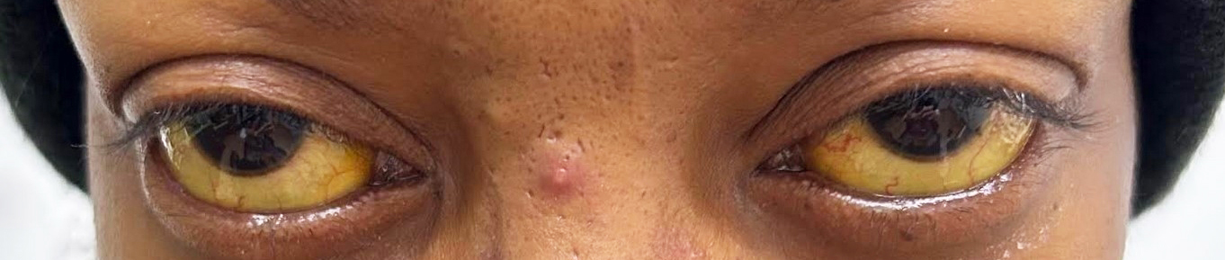
At this time, the patient was lost to follow-up, but she returned 3 months later with conjunctival icterus (Figure 4). The patient endorsed having anorexia and malaise but denied having stomach pain, changes in stool quality, or vomiting. After consultation with the clinic’s on-call doctor, the patient was advised to present to the emergency department with any worsening symptoms and to return the next day for an appointment with her primary care provider. The next day, her primary care provider placed a repeat referral to a gastroenterologist, ordered a computed tomography of the abdomen, and sent the patient to the laboratory for the previously discussed laboratory work along with repeat vitamin A levels.

Despite the patient’s reported compliance with oral vitamin A supplementation, the resulting laboratory work indicated lower vitamin A levels (10 mcg/dL) compared with several months prior. The patient also had low vitamin B1 levels (<7 nmol/L, reference range of 8-30 nmol/L). Despite high vitamin B12 (1753 pg/mL, reference range of 200-1100 pg/mL), low methylmalonic acid (56 nmol/L, reference range of 87-318 nmol/L), and normal serum folate levels (6.1 ng/mL, reference range of ≥5.5 ng/mL), the patient had high homocysteine levels (13.3 umol/L, reference range of <10.4 umol/L), which is indicative of a functional folate deficiency. After reviewing the laboratory results, the patient was called and agreed to be taken to the emergency department because of increasing symptoms of weakness and poor appetite.
The patient was admitted to the hospital where she was diagnosed with hypokalemia, hypomagnesemia, macrocytic anemia, malnutrition, and alcoholic cirrhosis of the liver. Over the course of the next 8 days, the patient was given IV potassium, IV folic acid, IV thiamine, and IV multivitamin in addition to medication to prevent hepatic encephalopathy and treat symptoms of alcohol withdrawal. Because of the patient’s weakness and lack of insight into her condition, she was advised to be placed in a skilled nursing facility; however, she left the hospital against medical advice to care for herself at home. At discharge, the patient was prescribed lipase-protease-amylase 12 000 units by mouth 3 times a day, diphenoxylate/atropine 2.5 mg/0.025 mg by mouth 4 times a day, famotidine 20 mg by mouth 2 times a day, folic acid 1 mg by mouth once a day, and rifaximin 600 mg by mouth 2 times a day.
The patient presented to the optometry department 4 days later. She presented with stable findings of moderate dry eyes and conjunctival icterus. She was advised to continue her dry eye treatment, continue taking 10 000 IU vitamin A per day until her next vitamin A assay, continue taking 1 mg folic acid per day, and start taking 100 mg vitamin B1 per day in addition to a daily multivitamin. The patient continued care with her primary care provider and a gastroenterologist and remained sober for the next 6 months. Her laboratory results at 6 months indicated normal levels of vitamin A, vitamin B12, and folate. Vitamin B1 was low but improved (7 nmol/L, reference range of 8-30 nmol/L). After the updated laboratory work, the patient’s primary care provider directed her to discontinue taking oral vitamin A but to continue taking folic acid, vitamin B1, and daily multivitamin supplements. At her concurrent eye examinations, the patient’s conjunctival icterus resolved, her Bitot spots resolved, and her symptoms of night blindness had significantly improved (Figure 5). Repeat fundus photos and optical coherence tomography studies demonstrated stability of fundus and optic nerve findings (Figure 6). The patient is receiving ongoing management for her dry eyes and monitoring for progression of her suspected bilateral nutritional optic neuropathy.
_temporal_and_(b)_nasal_conjunctiva_of_the_right_eye_6_months_a.tiff)
_right_and_(b)_left_eyes_and_optical_coherence_t.tiff)
DISCUSSION
Vitamin A is a fat-soluble essential vitamin stored primarily in the liver and found in a variety of foods, including fish, liver, eggs, dairy products, and many vegetables. It is a crucial vitamin for maintaining the health of the ocular surface and the retina, in addition to many other functions within the body, including immune function.1 Although vitamin A deficiency is uncommon in developed countries, it remains a public health concern in many developing countries because of inadequate diet, as it is the leading cause of preventable blindness in children.2 In developed countries, vitamin A deficiency can appear in patients with a highly restrictive diet, with liver disease, or with malabsorption because of a gastrointestinal disorder, pancreatic insufficiency, or gastric bypass operation.1,3
Vitamin A serves 2 critical functions in the eye. First, it is the precursor to rhodopsin, which is required for rods to conduct phototransduction. Because 11-cis-retinal in rhodopsin is derived from vitamin A, vitamin A must be continuously and readily available in the bloodstream for proper rod function. Second, it is required for maintenance of corneal and conjunctival epithelium. Therefore, vitamin A deficiency results in a host of adverse ocular manifestations, referred to as xerophthalmia. The earliest manifestation of xerophthalmia is night blindness, which has been shown to be both a sensitive and specific symptom of vitamin A deficiency because there is poor rod function with low serum levels of vitamin A and improved rod function with normal levels of vitamin A.4 It is likely that our patient had this as her symptoms of night blindness improved with vitamin A supplementation. Patients may also present with conjunctival xerosis due to the loss of goblet cells within the conjunctival epithelium, which can be observed as a dulled appearance of the conjunctival surface. The conjunctiva may also form Bitot spots: these spots are patches of keratinized epithelial cells on the conjunctiva that often appear foamy because of infiltration of gas-forming bacteria and are considered a pathognomonic sign of vitamin A deficiency.5 If xerophthalmia is prolonged, more severe ocular surface changes can manifest, including corneal xerosis, corneal ulceration, keratomalacia, and corneal scar formation. Although rare, patients with prolonged vitamin A deficiency may also develop a xerophthalmic fundus, characterized by deep white retinal lesions in the posterior pole or midperiphery, similar to drusenoid deposits in age-related macular degeneration.4
Patients with signs of xerophthalmia must be tested for serum vitamin A levels in addition to receiving traditional treatment for dry eyes. Although vitamin A deficiency is treated with supplementation, care should be taken to not overprescribe vitamin A, as it can also cause hepatotoxicity when taken in high doses.1 It is also critical to determine the cause of vitamin A deficiency with a thorough review of systems to determine any history of weight loss operation, restrictive eating habits, or gastrointestinal symptoms along with a comprehensive metabolic panel to determine whether liver disease is present. Patients with vitamin A deficiency can be monitored with routine serum vitamin A levels regularly every 2 to 4 months until the deficiency is resolved.4
Prolonged heavy alcohol consumption as seen with this patient can cause deficiency in many vitamins (including vitamins A, B1, B12, and folate) through inadequate intake as well as impaired absorption, storage, and metabolism of vitamins.6 Alcoholic liver disease further contributes to the imbalance of vitamins and other micronutrients, often causing abnormal accumulation of some nutrients, such as zinc, copper, and vitamin B12, while also causing depletion of other nutrients, such as folate and vitamin B1.6 These imbalances themselves have been shown to further contribute to disease morbidity with links to ascites, cirrhosis, and hepatic encephalopathy, and many studies encourage the monitoring and supplementation of multiple nutrients to improve organ function and patient outcomes.7–10
Liver cirrhosis can develop in longstanding alcoholic liver disease and is characterized by fibrosis of the liver and worsening liver function. Patients with compensated cirrhosis may be largely asymptomatic; therefore, liver disease is largely detected through laboratory work.11 Patients with decompensated cirrhosis, however, can present with various signs and symptoms, including jaundice, abdominal distention from ascites, vomiting or dark-colored stool from gastrointestinal bleeding, and confusion from hepatic encephalopathy.12 Decompensated cirrhosis indicates poor patient prognosis: 10-year survival drops from 47% in patients with compensated cirrhosis to 16% in patients with decompensated cirrhosis.11 Behavioral modification, including eliminating alcohol consumption, limiting sodium intake, and proper nutritional supplementation, is required to improve liver function; liver transplantation may be needed in some patients with severe decompensated cirrhosis.11
It is crucial to remember that because liver disease carries the risk of multiple nutritional deficiencies—and several of these deficiencies can cause different ocular manifestations—patients with liver disease need monitoring for a variety of ocular findings. Vitamin B1 deficiency can cause Wernicke’s encephalopathy, which has historically been characterized by the triad of encephalopathy, ophthalmoplegia, and gait ataxia; however, this triad has been shown to present in as few as 10% of cases.13 More recent recommendations suggest that patients presenting with any 2 of the following conditions should be presumed to have Wernicke’s encephalopathy: (1) nutritional deficiency, (2) ocular findings (including nystagmus or any other gaze abnormalities), (3) ataxia, or (4) mental status changes—using this criteria, our patient could be presumed to have had Wernicke’s encephalopathy when she presented to the emergency department because of her nutritional deficiencies and possible mental status changes.13 Vitamin B1 serum levels can be tested to determine the presence of a vitamin B1 deficiency. Vitamin B12 and folate work together in many biochemical functions—a deficiency in either or both vitamins can cause a nutritional optic neuropathy, characterized in later stages by reduced visual acuity (typically 20/50 or worse), very poor color vision, temporal optic nerve pallor, and central or cecocentral visual field deficits.14 However, with modern optical coherence tomography technology, it may now be possible to detect nutritional optic neuropathy in earlier stages by identifying the stereotypical findings of temporal thinning of the retinal nerve fiber layer and a diffusely thin ganglion cell complex.15 Serum levels of vitamin B12 and folate can be helpful in testing for deficiencies but do not always reflect tissue levels. Therefore, if serum B12 or folate is in the low or reference range and a deficiency is suspected, additional testing for methylmalonic acid and homocysteine should also be performed. These molecules are both intermediates in vitamin B12 and folate metabolism: both methylmalonic acid and homocysteine levels are elevated if the patient is functionally deficient in vitamin B12, but only homocysteine levels are elevated if the patient is functionally deficient in folate.16 Although deficiencies in other vitamins, such as vitamins C, D, and E, do not directly cause ocular manifestations, deficiencies in these nutrients may contribute to ocular morbidity, including subconjunctival hemorrhage (vitamin C), worsening dry eye (vitamins C, D, E), and an increased risk of various ocular diseases such as age-related macular degeneration (vitamins C, D, E) and glaucoma (vitamin D).16,17
Our patient was found to have deficiencies in vitamin A, vitamin B1, vitamin B12, folate, and magnesium. The cause of these deficiencies stems from alcohol abuse resulting in poor nutrition, pancreatic insufficiency, and liver cirrhosis. Management of all these conditions, including alcohol abstinence, proper nutrition, and management of pancreatic and liver disease, is required for prolonged resolution of her nutritional deficiencies. Additionally, monitoring for ocular signs of nutritional deficiencies is required to confirm stability of her ocular health. Management in collaboration with other health care professionals ensures this can happen efficiently and effectively for the patient.
CONCLUSION
Vitamin A deficiency can cause xerophthalmia, which can manifest in an array of signs including night blindness, corneal and conjunctival xerosis, Bitot spots, corneal ulceration, and keratomalacia. Recognizing these signs can be very helpful in identifying a deficiency in vitamin A as well as prompting tests of both serum levels of vitamin A and serum levels of other nutrients because the most common causes of vitamin A deficiency are also likely to cause deficiencies in other nutrients. Patients with alcoholism and alcoholic liver disease in particular have a significant risk of developing nutritional deficiencies and require collaborative management to monitor for liver and pancreatic disease, nutritional health, and associated ocular manifestations.